
Dental bone graft images:What are dental bone graft images and what do they typically show?
Q: What are dental bone graft images and what do they typically show?
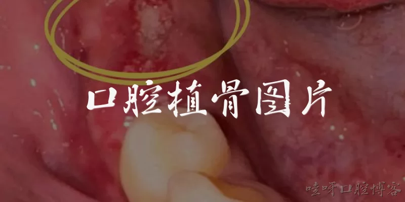
A: Dental bone graft images are radiographic or photographic records used to evaluate bone augmentation procedures. They typically show the grafted material, such as autograft, allograft, xenograft, or synthetic bone substitutes, placed in the alveolar ridge or extraction socket. Common imaging modalities include periapical radiographs, panoramic radiographs, cone beam computed tomography (CBCT), and intraoral photographs. According to the American Academy of Implant Dentistry, these images help clinicians assess graft integration, bone density, and healing over time. CBCT, in particular, provides three-dimensional visualization of bone volume, which is critical for implant site preparation and surgical planning, as noted in guidelines from the International Team for Implantology.
Q: When are dental bone graft images necessary for treatment planning?
A: Dental bone graft images are necessary when evaluating bone deficiencies before implant placement or after tooth extraction. According to the American Dental Association, radiographic imaging is essential for assessing bone quality and quantity, especially in cases of ridge augmentation or sinus lift procedures. The European Association for Osseointegration recommends CBCT for precise measurement of grafted bone volume and to avoid complications. Images are also required for monitoring healing at follow-up intervals. For example, a 2019 consensus report from the Osteology Foundation states that baseline and sequential imaging are vital to confirm graft incorporation and detect resorption. Without such images, clinicians cannot reliably determine implant feasibility or timing.
Q: What imaging modalities are commonly used for dental bone graft assessment?
A: Common modalities include periapical radiography, panoramic radiography, cone beam computed tomography (CBCT), and occasionally magnetic resonance imaging (MRI). Periapical and panoramic images provide two-dimensional overviews but have limitations in assessing buccolingual dimensions. CBCT is the preferred modality for three-dimensional evaluation of bone graft volume and density, as supported by the American Academy of Oral and Maxillofacial Radiology. A 2020 systematic review in the International Journal of Implant Dentistry found CBCT superior for detecting graft resorption and integration. MRI is rarely used due to cost and artifacts. Additionally, intraoral photographs and 3D scans can document soft tissue healing but do not replace radiographic assessment for bone graft evaluation.
Q: How do dental bone graft images help monitor graft healing and complications?
A: Dental bone graft images assist in monitoring healing by revealing changes in graft density, volume, and integration with host bone. According to a 2021 consensus report from the Academy of Osseointegration, sequential CBCT scans at 3, 6, and 12 months can detect resorption, infection, or graft failure. Radiographic signs of successful healing include increased radiopacity and trabecular bone formation. Complications such as graft exposure, infection, or non-union appear as radiolucent areas or loss of graft material. The American Association of Oral and Maxillofacial Surgeons emphasizes that imaging helps differentiate normal healing from pathology, guiding timely intervention. However, clinicians should minimize radiation exposure by using low-dose protocols and only when clinically justified.
Q: What do official guidelines say about interpreting dental bone graft images for implant success?
A: Official guidelines, such as those from the International Team for Implantology (ITI) and the American College of Prosthodontists, state that dental bone graft images must be interpreted by trained clinicians using standardized criteria. Key parameters include graft volume stability, bone density in Hounsfield units, and absence of radiolucent lines. The ITI’s 2018 consensus recommends that a minimum bone height and width, often 6 mm and 4 mm respectively, be confirmed on CBCT before implant placement. Additionally, the European Federation of Periodontology advises that images be correlated with clinical findings. These guidelines stress that imaging alone is insufficient; it must be part of a comprehensive evaluation to predict implant success and avoid complications.
Dialogue about
Common scenarios of "Dental bone graft images"
【Dentist】 Good morning, Mrs. Carter. I've reviewed your CBCT scan, and the bone loss in the lower left molar area is quite significant. We'll need a bone graft before placing an implant.
【Patient】 That sounds serious. Can you show me exactly what the bone graft images look like? I'm a visual person.
【Dentist】 Of course. Here on this cross-sectional slice, you can see the height and width of the remaining ridge. The graft material will fill this defect, and over time it will turn into your own bone.
【Patient】 So the white area in the image is the bone? And the dark part is where the bone is missing?
【Dentist】 Exactly. The bright white is dense bone, the grayish is less dense, and the black is air or soft tissue. The graft will be placed in that dark void.
【Patient】 How do you decide how much graft material to use? Is it based on those images?
【Dentist】 Yes, we measure the defect in three dimensions using software. The graft volume is calculated to restore the ridge to a shape that can support an implant of the right length and width.
【Patient】 Will I be able to see the graft on an X-ray after the surgery?
【Dentist】 Definitely. The graft material is radiopaque, so it will appear as a distinct granular or solid area on post-op images. Here's an example from a similar case.
【Patient】 Wow, that looks like a little block. Does it stay that shape forever?
【Dentist】 No, it gradually gets replaced by new bone. We take follow-up images at 3, 6, and 9 months to track the remodeling. The graft's appearance changes from granular to more bone-like.
【Patient】 What if the graft doesn't integrate well? Would the images show that?
【Dentist】 Yes, we'd see a radiolucent line around the graft or lack of density change. That would indicate failure, and we'd need to revise. But with proper technique, success rates are high.
【Patient】 Can you show me a before-and-after image of a successful graft? That would help me understand.
【Dentist】 Sure. On the left is the pre-op scan with the defect. On the right is 6 months after grafting. See how the ridge is now filled with a dense, homogeneous pattern?
【Patient】 That's amazing. So the new bone looks just like the original bone on the image?
【Dentist】 Almost. It may be slightly more radiopaque initially due to the graft particles, but eventually it becomes indistinguishable from native bone.
【Patient】 What kind of graft material do you use? Does it show up differently on images?
【Dentist】 We often use a mix of autogenous bone and a synthetic beta-tricalcium phosphate. Autogenous bone is slightly more radiopaque, but both are visible. The choice depends on the defect size.
【Patient】 So the images help you choose the material too?
【Dentist】 Yes, for larger defects we might use a block graft, which appears as a solid radiopaque block. For smaller ones, particulate graft looks like grains. The imaging guides that decision.
【Patient】 I feel much better seeing these images. It's like a roadmap for my jaw.
【Dentist】 Exactly. And we'll use these images to plan the implant placement later. The graft creates the foundation, and the images confirm it's ready.