Impacted wisdom tooth near the dental nerve:What does it mean when an impacted wisdom tooth is near the dental nerve?
Q: What does it mean when an impacted wisdom tooth is near the dental nerve?
A: When an impacted wisdom tooth lies near the inferior alveolar nerve (IAN), it means the tooth is positioned close to the main nerve canal in the lower jaw. This proximity raises the risk of nerve injury during extraction, potentially causing temporary or permanent numbness in the lip, chin, and gums. According to the American Association of Oral and Maxillofacial Surgeons (AAOMS) 2026 Clinical Practice Guidelines, preoperative imaging such as cone-beam CT (CBCT) is strongly recommended to assess the exact relationship between the tooth roots and the nerve. The guidelines note that while most patients recover fully, approximately 0.5–2% may experience persistent altered sensation. Therefore, careful evaluation and possibly a coronectomy—removing only the crown and leaving safe root fragments—may be advised to protect the nerve.
Q: How is an impacted wisdom tooth near the nerve diagnosed and assessed before surgery?
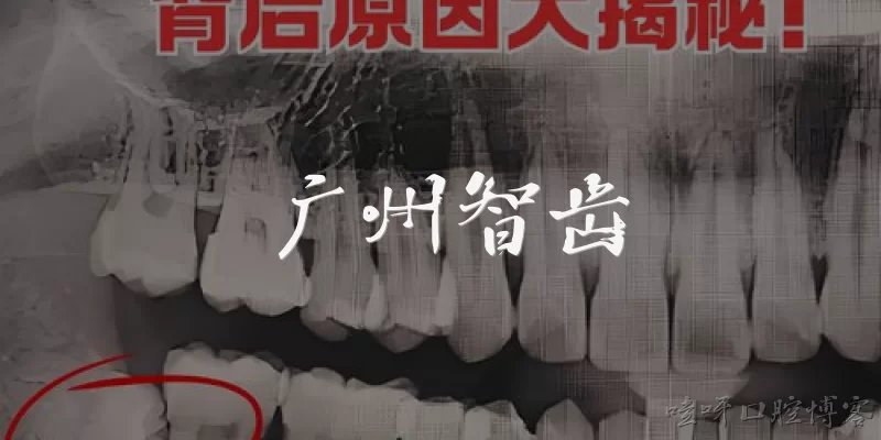
A: Diagnosis relies on clinical examination and advanced imaging. A panoramic X-ray is typically the first step, but it often cannot show the precise three-dimensional relationship between the tooth roots and the inferior alveolar nerve. The 2026 AAOMS guidelines recommend cone-beam computed tomography (CBCT) for any case where the roots appear close to or overlapping the nerve canal on a panoramic image. CBCT provides detailed cross-sectional views that reveal whether the nerve is buccal, lingual, or inferior to the roots, and whether there is cortical bone separation. Some clinicians also use risk assessment tools like the Rood and Shehab criteria. Based on these findings, the surgeon can decide whether to proceed with standard extraction, perform a coronectomy, or refer to a specialist. Proper imaging reduces the risk of unexpected nerve damage and helps in obtaining informed consent.
Q: What are the treatment options for an impacted wisdom tooth that is near the dental nerve?
A: Treatment options depend on symptoms, infection risk, and the exact nerve proximity. According to the 2026 American Dental Association (ADA) clinical update, if the tooth is asymptomatic and not causing harm, monitoring may be appropriate, especially if the patient has medical conditions that increase surgical risk. When removal is necessary, a standard extraction is possible if CBCT confirms a safe margin. However, for high-risk cases where the nerve is directly touching or enveloping the roots, a coronectomy is often preferred. This procedure removes the crown while leaving the roots undisturbed, reducing nerve injury risk by up to 90% according to AAOMS 2026 data. Alternatively, an orthodontic or surgical exposure may be considered if the tooth can be guided into function. In rare cases, a nerve-sparing approach using piezosurgery may also be used.
Q: What is the risk of nerve damage from removing an impacted wisdom tooth near the dental nerve, and how can it be managed?
A: The risk of injury to the inferior alveolar nerve (IAN) during removal of a lower impacted wisdom tooth near the nerve varies from 0.5% to 5% for temporary numbness and 0.1% to 2% for permanent altered sensation, according to the 2026 AAOMS Clinical Practice Guidelines. Risk is higher when radiographic signs show root narrowing, darkening, or deflection. To manage this, surgeons use CBCT to plan the approach. If the nerve is deemed at high risk, a coronectomy is recommended, which reduces permanent nerve injury to less than 0.1%. If nerve damage occurs, most patients recover within 6–12 months. Management includes observation, vitamin B complex supplements, and in severe cases, microsurgical nerve repair. The 2026 guidelines emphasize informed consent and a shared decision-making process between the patient and the oral surgeon.
Dialogue about
Common scenarios of "Impacted wisdom tooth near the dental nerve"
【Patient】 Hi, Doctor. I've been having this dull ache in the back of my mouth for weeks. My dentist said my wisdom tooth is impacted and near a nerve. I'm really worried.
【Doctor】 Hello, I understand your concern. An impacted wisdom tooth near the nerve can be tricky. Can you tell me more about your symptoms? Is the pain constant or does it come and go?
【Patient】 It comes and goes, but when it flares up, it's pretty bad. I also feel some numbness in my jaw sometimes. Could that be related?
【Doctor】 Yes, numbness could be a sign of nerve involvement. We need to assess the exact position of the tooth. Have you had any X-rays or a CT scan done?
【Patient】 I had a panoramic X-ray, and my dentist mentioned the tooth is very close to the inferior alveolar nerve. He referred me to you for further evaluation.
【Doctor】 Good. A panoramic X-ray gives a general idea, but for precise proximity, a CBCT scan is better. It provides 3D imaging to see exactly how close the tooth is to the nerve.
【Patient】 Is that necessary? I'm a bit concerned about the extra radiation and cost.
【Doctor】 I understand. The radiation dose from a CBCT is low and focused. It's crucial to avoid nerve damage during extraction. The cost is worth it to prevent potential permanent numbness.
【Patient】 Okay, that makes sense. What are the risks if I decide not to remove the tooth?
【Doctor】 If left untreated, the impacted tooth can cause infection, cyst formation, damage to adjacent teeth, and worsening pain. The nerve might also be affected over time.
【Patient】 And if I do remove it, what are the chances of nerve damage?
【Doctor】 With careful planning using CBCT, the risk is low but not zero. Possible complications include temporary or permanent numbness of the lip, chin, and gums. We'll discuss all risks and benefits before proceeding.
【Patient】 That sounds scary. Is there any alternative to surgery?
【Doctor】 In some cases, we might consider a coronectomy, where we remove only the crown and leave the roots to avoid nerve injury. But it depends on the tooth's condition and position.
【Patient】 What's the recovery like for a regular extraction?
【Doctor】 Typically, swelling and discomfort for a few days, managed with ice packs and pain meds. You'll need to eat soft foods and avoid strenuous activity. Full recovery takes about a week or two.
【Patient】 And for coronectomy? Is it different?
【Doctor】 Similar, but there's a chance the root may need removal later if it causes problems. We monitor it with X-rays.
【Patient】 I see. I think I'll go ahead with the CBCT scan. Can you schedule that for me?
【Doctor】 Of course. I'll write you a referral. After the scan, we'll review the images together and decide on the best approach. Any other questions?