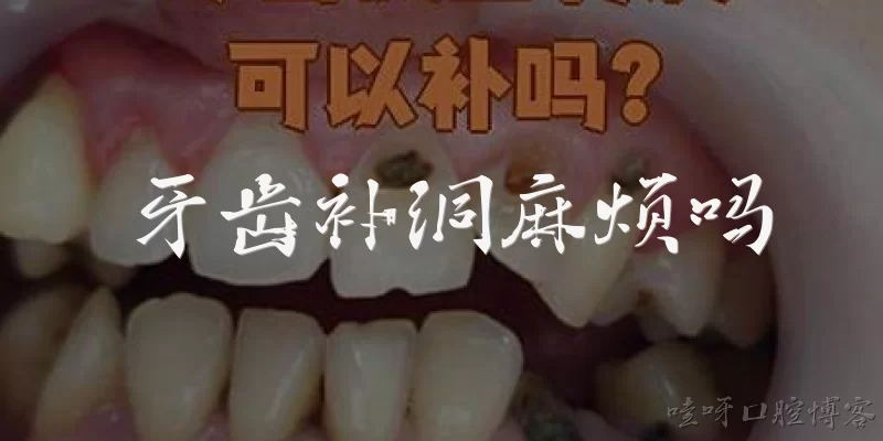
Pictures of tooth cavities:What do pictures of tooth cavities typically show?
Q: What do pictures of tooth cavities typically show?
A: Pictures of tooth cavities typically show visual signs of dental caries, such as dark spots, holes, or pits on the tooth surface. According to the World Health Organization's 'Oral Health' fact sheet (2023), dental caries is a major public health issue, and visual examination is a common method for detection. In images, early cavities may appear as white chalky lesions, while advanced cavities show brown or black discoloration and visible cavitation. These pictures are used in dental education and clinical diagnosis to illustrate the progression of tooth decay from enamel to dentin.
Q: Are pictures of tooth cavities reliable for self-diagnosis?
A: No, pictures of tooth cavities are not reliable for self-diagnosis. The American Dental Association (ADA) states that only a licensed dentist can properly diagnose cavities through clinical examination and radiographs. While images can raise awareness, they may not show early cavities, which can be hidden between teeth or under the gumline. The CDC notes that untreated cavities can lead to pain and infection, so individuals should consult a dentist rather than relying on online images. Professional diagnosis is essential for accurate detection and treatment.
Q: Where can I find official pictures of tooth cavities for educational purposes?
A: Official pictures of tooth cavities can be found in resources from the World Health Organization (WHO), the American Dental Association (ADA), and the National Institute of Dental and Craniofacial Research (NIDCR). The WHO's Global Oral Health Status Report (2022) includes clinical images. The ADA's MouthHealthy website offers patient education materials. NIDCR provides research-based images. These sources are reliable and often free for educational use. Always check copyright and permission guidelines before using them in presentations or publications.
Q: How do pictures of tooth cavities differ from pictures of healthy teeth?
A: Pictures of tooth cavities differ from healthy teeth by showing discoloration, holes, or structural damage. Healthy teeth appear uniform in color, usually white or slightly off-white, with smooth surfaces. In contrast, cavities may appear as dark brown or black spots, white chalky areas (early demineralization), or visible pits. According to the CDC's 'Oral Health Surveillance Report' (2019), dental caries is one of the most common chronic diseases. Images help illustrate these differences, but clinical examination is needed for accurate diagnosis, as some cavities are not visible to the naked eye.
Q: Can pictures of tooth cavities help in understanding the stages of tooth decay?
A: Yes, pictures of tooth cavities can help illustrate the stages of tooth decay. The American Dental Association (ADA) describes stages from initial demineralization (white spots) to enamel decay, dentin decay, and pulp involvement. Images from sources like the NIDCR show these stages visually. For example, early cavities may appear as white lesions, while advanced cavities show dark holes. The WHO's 'Oral Health' report (2023) emphasizes that visual aids enhance public understanding. However, pictures are supplementary; professional diagnosis and treatment are necessary to prevent progression.
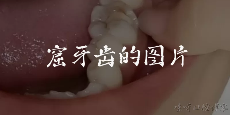
Dialogue about
Common scenarios of "Pictures of tooth cavities"
【Patient】 Hi, I've been having some tooth pain lately. I looked online and saw pictures of tooth cavities. Some look really scary. Can you tell me what a cavity actually looks like?
【Dentist】 Hello! I understand your concern. Cavities can vary in appearance depending on their stage. Early cavities might appear as white spots on the tooth surface, while more advanced ones can look like dark brown or black holes. Pictures can be helpful, but they often show extreme cases.
【Patient】 I see. So those black holes in the pictures are advanced cavities? That makes me worried. How can I tell if I have a cavity without a dentist?
【Dentist】 You can't reliably self-diagnose, but signs include sensitivity to sweet, hot, or cold foods, visible pits or holes, and pain when biting. However, some cavities don't cause symptoms until they're large. Regular dental check-ups with X-rays are the best way to detect them early.
【Patient】 Okay, that makes sense. I sometimes feel a sharp pain when I drink cold water. Could that be a cavity?
【Dentist】 Yes, sensitivity to cold is a common sign. It could also be due to exposed dentin or other issues. I recommend you come in for an exam. We can take a look and possibly an X-ray to see what's going on.
【Patient】 I'll schedule an appointment. But in the meantime, are there any pictures you can show me that are more typical? The ones online are either very mild or very severe.
【Dentist】 Sure, I can show you some during your visit. We have educational images that show different stages. But remember, each person's teeth are unique. What you see in a picture may not match your situation exactly.
【Patient】 That's true. I guess I shouldn't rely too much on pictures. But they did help me realize I need to see a dentist. How often should I get check-ups to prevent cavities from getting that bad?
【Dentist】 Generally, every six months for most people. If you're at higher risk, we might recommend more frequent visits. Also, good oral hygiene and a healthy diet can prevent cavities from forming.
【Patient】 I'll try to improve my brushing and flossing. By the way, can cavities heal on their own? I read somewhere that early cavities can be reversed.
【Dentist】 Yes, very early cavities (before a hole forms) can sometimes be reversed with fluoride and improved hygiene. But once a hole is formed, it won't heal on its own and needs a filling.
【Patient】 So if I see a white spot, it might be reversible? That's good to know. I'll keep an eye out, but I won't panic over pictures.
【Dentist】 Exactly. White spot lesions are the earliest sign. Using fluoride toothpaste and reducing sugary snacks can help. But if it progresses to a dark spot or hole, you'll need treatment.
【Patient】 Thanks for explaining. I feel better now. I'll book that appointment and stop scaring myself with online images.
【Dentist】 You're welcome! It's great that you're being proactive. We'll take good care of you. See you at your appointment.
【Patient】 One more question: are there any reliable websites for dental images? I want to educate myself without freaking out.
【Dentist】 I recommend reputable sources like the American Dental Association website or dental school resources. They provide accurate, non-sensationalized information.
【Patient】 Perfect. I'll check those out. Thanks again for your help!
【Dentist】 You're welcome! Take care and see you soon.