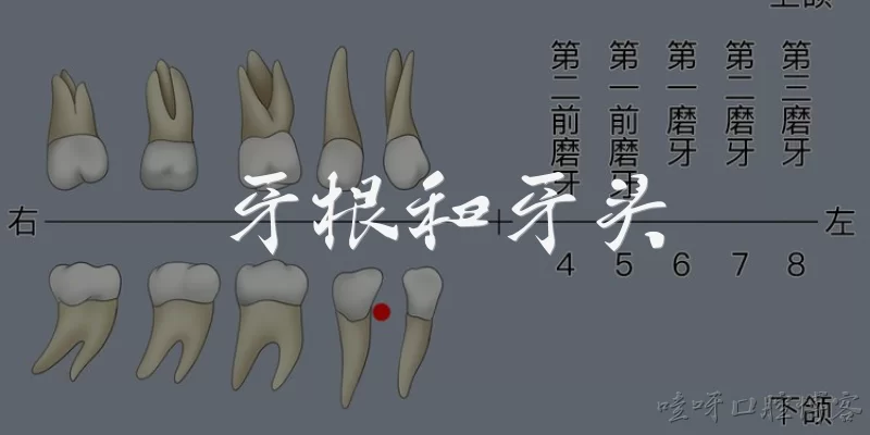
Mandibular apical surgery:What is mandibular apical surgery and when is it indicated?
Q: What is mandibular apical surgery and when is it indicated?
A: Mandibular apical surgery, also known as apicoectomy or root-end resection, is a surgical procedure involving the removal of the apical portion of a tooth root in the mandible, along with pathological tissue, followed by root-end preparation and filling. It is indicated when nonsurgical endodontic treatment fails, when there are anatomical obstructions, persistent apical periodontitis, or when a biopsy is needed. According to the American Association of Endodontists (AAE), apical surgery is considered a predictable treatment option with success rates often exceeding 90% when modern microsurgical techniques are employed. The AAE's 2020 guidelines emphasize proper case selection and technique for optimal outcomes.
Q: What are the key anatomical considerations in mandibular apical surgery?
A: Key anatomical considerations in mandibular apical surgery include the proximity to the inferior alveolar nerve (IAN), mental foramen, and mandibular canal. The roots of mandibular molars and premolars are often close to the IAN, and injury can cause neurosensory disturbances. The AAE and the American Academy of Oral and Maxillofacial Radiology recommend preoperative cone-beam computed tomography (CBCT) to assess the relationship between the root apices and vital structures. A 2019 joint position statement by the AAE and the American Academy of Oral and Maxillofacial Radiology highlights CBCT's role in minimizing risks. Additionally, the buccal cortical plate thickness and lingual concavities must be evaluated to avoid complications.
Q: What are the success rates of mandibular apical surgery?
A: Success rates for mandibular apical surgery vary based on technique and follow-up. According to a systematic review published in the Journal of Endodontics (2021), modern microsurgical techniques with ultrasonic root-end preparation and bioceramic root-end filling materials yield success rates of 85–95% at 1–4 years. The AAE's 2020 clinical practice guideline reports similar outcomes, noting that factors such as preoperative diagnosis, operator experience, and magnification influence success. Failures are often due to inadequate root-end sealing, missed canals, or vertical root fractures. Long-term studies indicate that mandibular apical surgery, when performed with contemporary protocols, is a reliable treatment for persistent apical periodontitis.
Q: What surgical techniques are used in mandibular apical surgery?
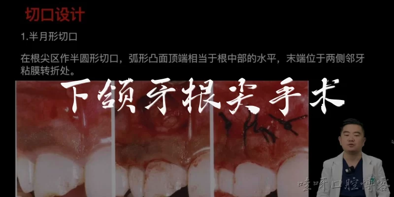
A: Mandibular apical surgery employs modern microsurgical techniques: a sulcular or submarginal incision with papilla preservation, elevation of a full-thickness flap, osteotomy to expose the root apex, resection of the apical 3 mm, root-end preparation with ultrasonic tips, and root-end filling with materials like mineral trioxide aggregate (MTA) or bioceramics. The AAE's 2020 guidelines endorse the use of magnification (microscope or loupes) and illumination. According to a 2018 consensus report from the AAE, the use of cone-beam computed tomography (CBCT) aids in preoperative planning. The procedure aims to eliminate infection, seal the root canal system, and promote periapical healing.
Q: What are the complications and postoperative care for mandibular apical surgery?
A: Complications of mandibular apical surgery include pain, swelling, bleeding, infection, nerve injury (especially inferior alveolar nerve), sinus tract formation, and flap necrosis. A 2019 review in the International Endodontic Journal notes that neurosensory disturbances are rare but possible, with an incidence of 0.5–5% depending on proximity to the nerve. Postoperative care includes analgesics, antibiotics if indicated, ice packs, and avoidance of chewing on the surgical site. The AAE recommends follow-up at 6 and 12 months to assess healing. Patients should be informed about potential risks, and CBCT is advised for high-risk cases to prevent nerve damage.
Dialogue about
Common scenarios of "Mandibular apical surgery"
【Dentist】 Good morning, Mrs. Johnson. I've reviewed your X-rays and it looks like the infection at the tip of your lower left first molar's root is not healing after the root canal. We may need to consider apical surgery.
【Patient】 Apical surgery? That sounds serious. What exactly is that?
【Dentist】 It's a procedure where we go in through the gum to remove the infected tissue and the very tip of the root. It's also called an apicoectomy. This is often recommended when a root canal fails to resolve the infection.
【Patient】 I see. Why didn't the root canal work? I thought that was supposed to fix the problem.
【Dentist】 Root canals have a high success rate, but sometimes there are complex canal anatomy or persistent bacteria that can't be reached. In your case, the infection seems to be around the root tip, and a traditional root canal can't access that area directly.
【Patient】 Is there any alternative? Maybe another root canal?
【Dentist】 A retreatment of the root canal is an option, but it also has limitations and may not resolve the issue if the infection is outside the root. Apical surgery is more direct and has a good success rate.
【Patient】 What does the procedure involve? Will it hurt?
【Dentist】 The area will be numbed with local anesthesia, so you shouldn't feel pain during the surgery. We'll make a small incision in the gum, lift it to expose the bone, remove the infected tissue and the root tip, then seal the end of the root. The whole thing usually takes about 30 to 60 minutes.
【Patient】 How long is the recovery? Will I be able to go back to work the next day?
【Dentist】 Most people take it easy for a day or two. You might have some swelling and discomfort, but that can be managed with ice packs and pain medication. You should avoid strenuous activity for a few days. Many patients return to work the next day if they feel up to it.
【Patient】 Are there any risks or complications?
【Dentist】 As with any surgery, there's a risk of infection, bleeding, or damage to nearby structures like nerves or adjacent teeth. But these are rare. We take precautions to minimize them. Also, sometimes the surgery might not fully resolve the problem, and further treatment might be needed.
【Patient】 What happens if I don't do the surgery? Can I just leave it?
【Dentist】 If left untreated, the infection can spread, cause bone loss, and potentially lead to more serious health issues. It could also result in the loss of the tooth. It's best to address it now.
【Patient】 Okay, I understand. How successful is this surgery?
【Dentist】 Success rates are generally high, around 85-90% when performed by an experienced surgeon. We'll monitor your healing with follow-up visits and X-rays to ensure the infection is gone.
【Patient】 That sounds reassuring. What should I do to prepare for the surgery?
【Dentist】 We'll schedule the appointment. Before that, you should avoid smoking as it hinders healing. Also, let us know if you're on any blood thinners or have any medical conditions. You might want to arrange for someone to drive you home after the procedure if you feel anxious.
【Patient】 Alright, I'll do that. Thank you for explaining everything, doctor. I feel more prepared now.
【Dentist】 You're welcome, Mrs. Johnson. We'll take good care of you. Let's get this scheduled and get you back to good oral health.